Describe ARMD
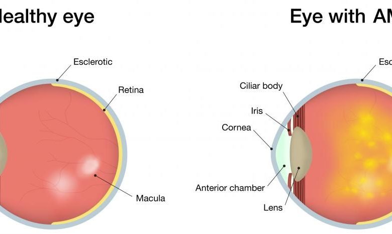
Age-related macular degeneration (ARMD) is an acquired retinal degeneration that considerably reduces central vision as a result of drusen and retinal pigment epithelium abnormalities as well as neovascular derangements (choroidal neovascular membrane formation). Serous fluid, subretinal fibrosis, and bleeding behind the retinal pigment epithelium (RPE) or beneath the retina may all be symptoms of an advanced illness. There is an urgent demand for innovative drugs due to recently implicated biochemical pathways and the absence of effective treatments for the majority of disorders (i.e., dry ARMD).
Also Read: Before And After An Eye Lift: The Transformation Is Incredible!
AMD Signs and Symptoms
Dry AMD
Most patients still have enough vision to read and drive despite the gradual, painless loss of central vision. Scotomas, or central blind patches, typically appear late in the course of the illness and can occasionally be severe. Typically, symptoms are bilateral.
The following alterations are caused by funduscopic changes:
- Retinal pigment epithelium changes
- Drusen
- Areas of chorioretinal atrophy
Wet AMD
Wet AMD is more common when vision loss occurs suddenly, typically spanning days to weeks. Visual distortion, such as a central blind spot (scotoma) or curving of straight lines, is frequently the initial symptom (metamorphopsia).
The patient runs the danger of become legally blind or having vision of 20/200 or worse in the affected eye. if AMD is not treated. Normally, colour and peripheral vision remain unaffected. Wet AMD symptoms are frequently unilateral since it typically only affects one eye at a time.
The following alterations are caused by funduscopic changes:
- Localized retinal elevation caused by subretinal fluid
- Retinal swelling
- Discoloration under the macula that is gray-green
- Macula exudates or exudates nearby
- Retinal pigment epithelium separation (visible as an area of retinal elevation)
- Subretinal bleeding in or near the macula.
Do I have an AMD risk?
As you age, your chance of developing AMD rises. AMD is more common in people over the age of 55. A person is also more likely to get AMD if they:
- History of AMD in your family
- Are White Smokers
Regular eye exams are crucial if you have AMD because of your age, family history, or other causes. How frequently should you have eye exams? Ask our best eye doctor in west Delhi. Don’t wait for your eyesight to start changing because early AMD doesn’t have any symptoms!
How may my risk of AMD be reduced?
By making these healthy decisions, research suggests that you may be able to reduce your risk of AMD (or slow vision loss from AMD):
- Don’t start smoking or stop smoking.
- Participate in regular exercise
- Maintain normal cholesterol and blood pressure levels.
- Consume nutritious foods like fish and leafy greens.
Diagnosis & Tests
When you see our best eye doctor (at bharti eye foundation) for a standard eye exam and have your eyes dilated, they can screen you for age-related macular degeneration. An early diagnosis will allow you to begin therapy, which could delay certain symptoms or lessen their severity.
Tests
Your retina, a layer of tissue in the back of your eye that processes light, will be examined along with your eyesight and eye pressure. They will look for drusen, which are little yellow deposits, under the retina. It is a normal first sign of the illness. In addition, your doctor might ask you to look at an Amsler grid, a pattern of straight lines that resembles a checkerboard.
If you observe that some of the lines are missing or have a wavelike appearance, macular degeneration may be present.
Tests If our doctor suspects you have age-related macular degeneration, they might recommend one or both of the following tests:
- Optical coherence tomography (OCT). A magnified 3D image of your retina may be seen in this unique photograph. Using this technique, your doctor can determine whether the retinal layers are misaligned. Additionally, whether you underwent laser or injectable therapy, they can determine whether the edema is becoming better or worse.
- Imaging with fluorescein. In this treatment, a dye is injected into a vein in your arm by your doctor.
They capture images as the dye enters your eye and travels through the retina’s blood vessels. Images of the macula, a small region near the center of your retina, will reveal new veins or vessels that are leaking blood or fluid.
Treatment & Prevention
Age-related macular degeneration (AMD) has no known cure, however, treatment can delay the progression of the condition and prevent serious vision loss. Consult our best eye doctor for advice on how to treat your illness.
Your Available Treatments
- Anti-angiogenic drugs. Your doctor administers these medications intravenously. They halt the growth of brand-new blood vessels and stop leakage from aberrant blood vessels that lead to wet macular degeneration.
- Some persons with AMD who use these medications have been able to restore their eyesight. On subsequent appointments, you’ll probably need to have the same treatment again.
- Photodynamic laser therapy: It’s a two-step procedure that damages your abnormal blood vessels with a light-sensitive medication.
- Your doctor injects a medication into your bloodstream, and the erratic blood vessels in your eye absorb it. The drug is then activated and the aberrant blood vessels are damaged using a laser.
Low vision aids: You can purchase tools with unique lenses or electrical systems that magnify images of adjacent objects.
These comprise:
- Magnifiers for reading (handheld or electronic)
- Glasses with unique lenses
- Binocular-equipped eyewear
- Electronic spectacles
- Apps for phones Products with large print (phones, clocks, print readers)
Next Steps for Macular Degeneration
Some AMD sufferers might transform from the dry to the wet type.
It’s crucial to keep an eye on your vision if you currently have dry form. Every week, check your vision, focusing on each eye independently. Use an Amsler Grid Chart as directed; it can be displayed on a tablet or computer, or it can be placed on your refrigerator. If something changes, inform your doctor.
Even after treatment, if you have a wet form of macular degeneration, you should test your eyesight to see if any blind spots reappear or grow larger. Months or even years after receiving injections or laser therapy, new blood vessels may start to appear.
If you just have AMD in one eye, your doctor will do routine eye exams in that eye to look for any symptoms of emerging issues.
More: Key difference to keep an eye on between PHP and Node.JS in this 2021
What are the prospects?
Age-related macular degeneration seldom causes complete blindness in people. Even if you have advanced AMD, you will still be able to see items to the side and out of your field of sight, while having impaired central vision. And many of your regular daily tasks will still be possible for you to complete.
Your central vision could become less than 20/200 in both eyes if you have wet or dry AMD that is severe. You will have peripheral vision, but your vision issues meet the criteria for legal blindness.
You can maintain the majority of your eyesight if you have the dry form of AMD, which is far more prevalent.
Wet AMD can occasionally return even after you receive therapy for it. Follow your doctor’s advice and perform regular vision tests. The appropriate course of treatment can enhance your vision in addition to slowing your vision loss.
This extensive guide about age related macular degeneration also known as armed or amd eye is posted here.


https://vyzov-santehnika-na-dom.ru.
Профессиональный сервисный центр по ремонту бытовой техники с выездом на дом.
Мы предлагаем: сервисные центры по ремонту техники в мск
Наши мастера оперативно устранят неисправности вашего устройства в сервисе или с выездом на дом!
Your point of view caught my eye and was very interesting. Thanks. I have a question for you.
I’ve been taking cornbread hemp full-spectrum cbd gummies concerning a while at once, and they’ve sincerely been a game-changer for stress and sleep. The excellent part? No grogginess in the morning just a undisturbed, relaxed impression rather than bed. Gain, they have knowledge of outstanding, unequivalent to some other supplements I’ve tried. I was skeptical at elementary, but after daily using them, I can decidedly say they avoid with unwinding after a extended day. If you’re looking for a ordinary personality to the sniffles without any weird side effects, CBD gummies are benefit trying. Honest get sure you fathom a grade brand with third-party testing!